 Total Heart Sim
The ICE views, around the clock.
Intracardiac echo views are taught as positions on a clock: the home view, facing the tricuspid valve, is 12 o'clock, and rotating the catheter clockwise moves the hour forward. Each view below shows the ICE image beside the beating 3D heart, with the imaging plane cutting through it.
- Home viewHome: 12 o'clock
- RV outflow and pulmonary valve12 o'clock, no rotation
- Aortic valveAbout 12 o'clock (10° clockwise)
- Left atrium and mitral valveAbout 2 o'clock (50° clockwise)
- Left atrial appendageAbout 3 o'clock (80° clockwise)
- Left pulmonary veinsAbout 3 o'clock (90° clockwise)
- Right pulmonary veinsAbout 5 o'clock (140° clockwise)
- Septal view and fossa ovalisAbout 4 o'clock (108° clockwise)
- Cavotricuspid isthmus12 o'clock, no rotation
- LV short axis and pericardiumAbout 2 o'clock (60° clockwise)
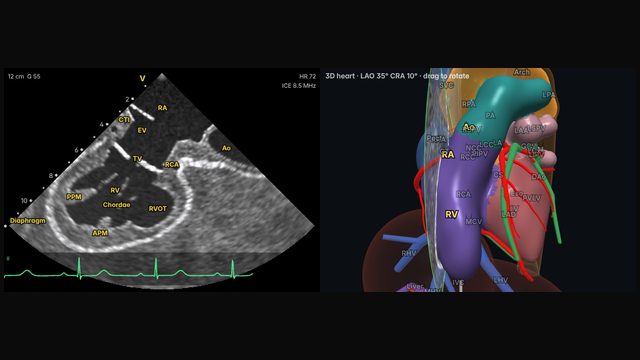
Home viewMid right atrium, neutral catheter: the tricuspid valve and RV. Every other view starts here.
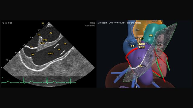
RV outflow and pulmonary valveThe outflow of the right ventricle up to the pulmonary valve: key for outflow tract VT and PVCs.
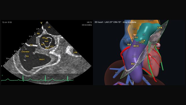
Aortic valveA small clockwise turn from home brings in the aortic root and its cusps.
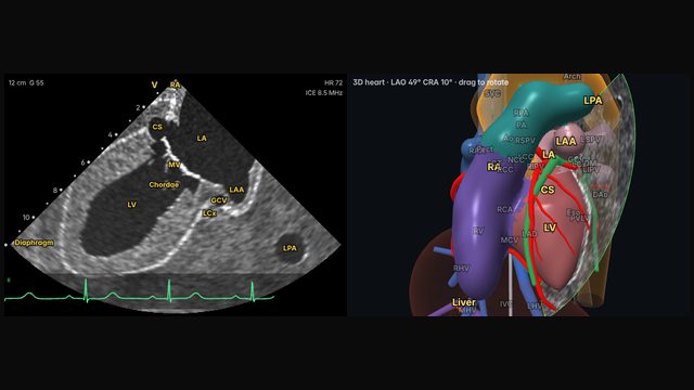
Left atrium and mitral valveFurther clockwise, across the septum: the left atrium with the mitral valve beyond.
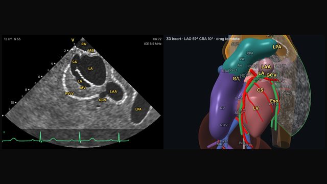
Left atrial appendageThe appendage at the edge of the left atrium: far from the catheter in the RA, which limits detail.
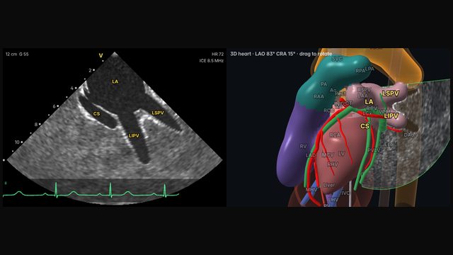
Left pulmonary veinsThe left superior and inferior veins entering the left atrium: a core view for AF ablation.
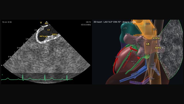
Right pulmonary veinsThe right-sided veins, close behind the septum: more rotation and deflection than the left veins.
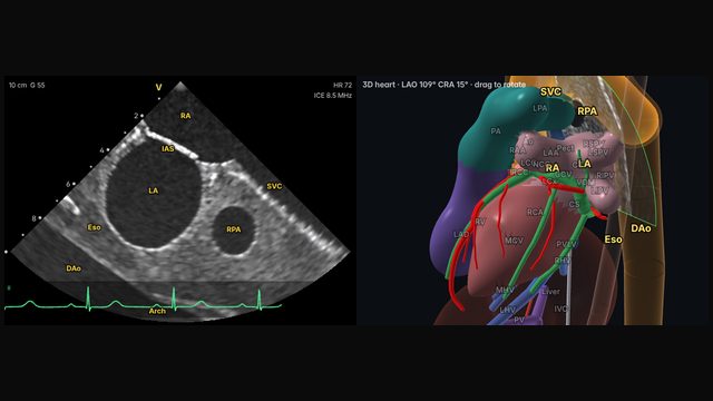
Septal view and fossa ovalisThe interatrial septum and fossa ovalis in the near field: the view for transseptal puncture.
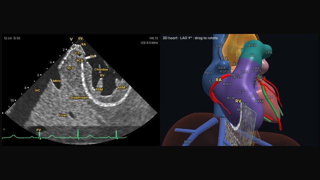
Cavotricuspid isthmusThe strip between the IVC and the tricuspid valve: the target for typical flutter ablation.
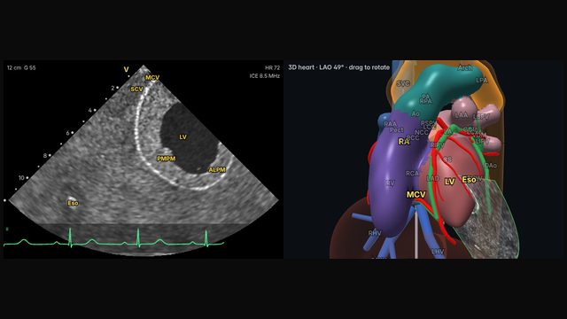
LV short axis and pericardiumThe left ventricle in cross-section with its papillary muscles, and the pericardial space around it.
Steer to every view yourself, with the 3D heart beside the echo.
Educational content; not medical advice. Positions are typical teaching values in the simulator's model heart; real anatomy and catheters vary.