 Total Heart Sim
What you see
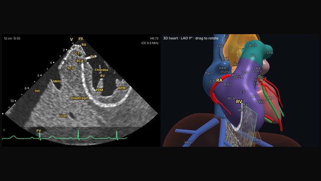
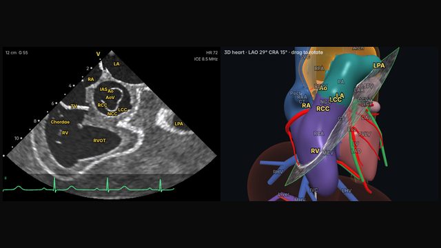
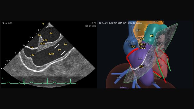
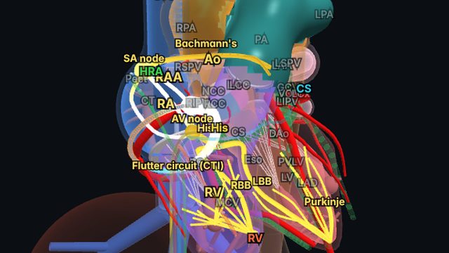
With the catheter in the mid right atrium and the handle in neutral, the imaging plane faces the tricuspid valve. The right atrium fills the near field, the tricuspid valve sits in the middle, and the right ventricle, its inflow and the RV outflow tract open out beyond it. The aortic root is often caught obliquely at the edge of the sector.
Why it matters
The home view is the reference point for everything else. Operators return to it whenever they lose orientation, and describe the other views by how far the catheter is rotated, deflected or advanced from here. On this site and in many teaching programs that rotation is described as a clock: home is 12 o'clock, and clockwise rotation moves the hour forward.
It is also the first look for the tricuspid valve, the right ventricle and the pericardial space around the RV.
How to get there
Home: 12 o'clock
The starting point: the catheter in the mid right atrium, handle neutral, no deflection, facing the tricuspid valve.
In the simulator:
- Place the catheter in the mid right atrium.
- Keep the handle neutral: no rotation, no deflection.
- The tricuspid valve should face the transducer, with the RV beyond it.
Clinical uses
- Orientation and a safe return point during any procedure.
- Tricuspid valve and RV assessment.
- A first look for pericardial effusion around the right heart.
Frequently asked questions
How do you get the home view view on ICE?
The starting point: the catheter in the mid right atrium, handle neutral, no deflection, facing the tricuspid valve. Small adjustments either way are normal: anatomy and catheters vary.
References
- Enriquez A, Saenz LC, Rosso R, et al. Use of intracardiac echocardiography in interventional cardiology: working with the anatomy rather than fighting it. Circulation. 2018;137:2278–2294.
- Haloot J, Schurmann P. Electrophysiology laboratory survival guide part 2: introduction to intracardiac echocardiography in the electrophysiology laboratory. US Cardiology Review. 2026;20:e16.
- Alkhouli M, Hijazi ZM, Holmes DR Jr, Rihal CS, Wiegers SE. Intracardiac echocardiography in structural heart disease interventions. JACC Cardiovasc Interv. 2018;11(21):2133–2147.
Educational content; not medical advice. Rotation and deflection values are typical teaching values in the simulator's model heart; real anatomy and catheters vary.