 Total Heart Sim
What you see
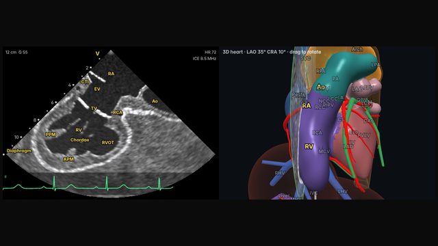
Low in the right atrium, the cavotricuspid isthmus runs from the inferior vena cava and Eustachian ridge to the tricuspid annulus. The view shows its length and shape: some isthmuses are short and flat, others long, pouched (a sub-Eustachian recess) or crossed by a prominent Eustachian ridge.
Why it matters
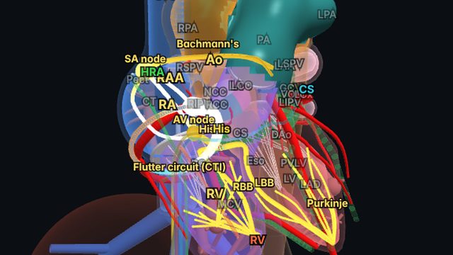
Typical atrial flutter depends on the isthmus, and a complete ablation line across it cures the flutter. Difficult anatomy is a common reason lines are hard to complete. Seeing the ridge, any pouch and the ablation catheter's contact directly helps place lesions where they count.
How to get there
12 o'clock, no rotation
From the low right atrium, anterior deflection lays the isthmus out between the IVC and Eustachian ridge and the tricuspid annulus.
In the simulator:
- Start from the home view (mid right atrium, neutral).
- Deflect anteriorly (about 60° on the A/P dial).
Clinical uses
- Typical flutter ablation: isthmus anatomy and catheter contact.
- Identifying pouches and a prominent Eustachian ridge.
Frequently asked questions
How do you get the cavotricuspid isthmus view on ICE?
From the low right atrium, anterior deflection lays the isthmus out between the IVC and Eustachian ridge and the tricuspid annulus. In the Total Heart Sim model heart: Deflect anteriorly (about 60° on the A/P dial). Small adjustments either way are normal: anatomy and catheters vary.
References
- Enriquez A, Saenz LC, Rosso R, et al. Use of intracardiac echocardiography in interventional cardiology: working with the anatomy rather than fighting it. Circulation. 2018;137:2278–2294.
- Haloot J, Schurmann P. Electrophysiology laboratory survival guide part 2: introduction to intracardiac echocardiography in the electrophysiology laboratory. US Cardiology Review. 2026;20:e16.
Educational content; not medical advice. Rotation and deflection values are typical teaching values in the simulator's model heart; real anatomy and catheters vary.