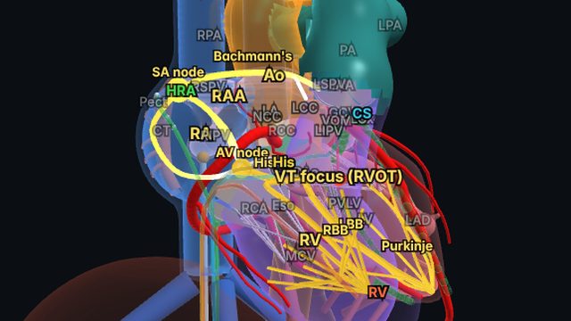
 Total Heart Sim
What you see
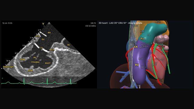
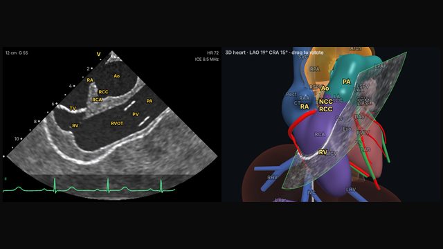
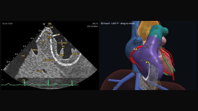
The left ventricle appears in cross-section with its anterolateral and posteromedial papillary muscles, surrounded by the pericardium. Operators commonly obtain the cleanest short axis by advancing the catheter into the right ventricle; from the right atrium, as in the simulator, a similar view comes from rotation with anterior and leftward deflection.
Why it matters
Pericardial effusion is one of the most important complications to catch early during ablation, and ICE can show a new echo-free space around the heart before blood pressure falls. The same view guides ablation near the papillary muscles, a site of origin for some ventricular arrhythmias.
How to get there
About 2 o'clock (60° clockwise)
Operators usually advance across the tricuspid valve into the RV and deflect so the plane crosses the LV in short axis; from the right atrium, a similar view comes from rotation with anterior and leftward deflection.
In the simulator:
- Start from the home view (mid right atrium, neutral).
- Withdraw the catheter about 1.0 cm, lower in the right atrium.
- Rotate clockwise about 60°, to about 2 o'clock.
- Deflect anteriorly (about 40° on the A/P dial).
- Tilt left (about 40° on the R/L dial).
Clinical uses
- Early detection of pericardial effusion during procedures.
- Papillary muscle arrhythmia ablation: catheter position and contact.
- LV function at a glance.
Frequently asked questions
How do you get the lv short axis and pericardium view on ICE?
Operators usually advance across the tricuspid valve into the RV and deflect so the plane crosses the LV in short axis; from the right atrium, a similar view comes from rotation with anterior and leftward deflection. In the Total Heart Sim model heart: Withdraw the catheter about 1.0 cm, lower in the right atrium. Rotate clockwise about 60°, to about 2 o'clock. Deflect anteriorly (about 40° on the A/P dial). Tilt left (about 40° on the R/L dial). Small adjustments either way are normal: anatomy and catheters vary.
References
- Enriquez A, Saenz LC, Rosso R, et al. Use of intracardiac echocardiography in interventional cardiology: working with the anatomy rather than fighting it. Circulation. 2018;137:2278–2294.
- Haloot J, Schurmann P. Electrophysiology laboratory survival guide part 2: introduction to intracardiac echocardiography in the electrophysiology laboratory. US Cardiology Review. 2026;20:e16.
Educational content; not medical advice. Rotation and deflection values are typical teaching values in the simulator's model heart; real anatomy and catheters vary.