 Total Heart Sim
What you see
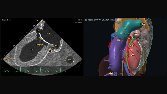
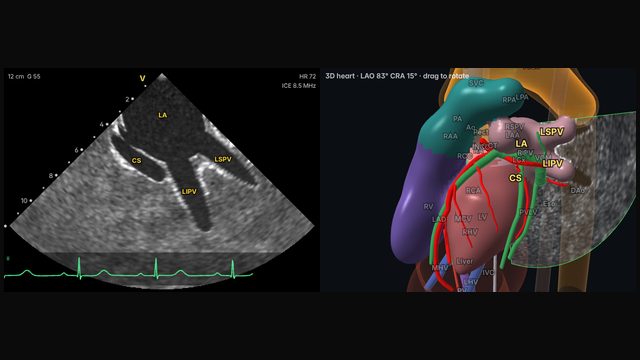
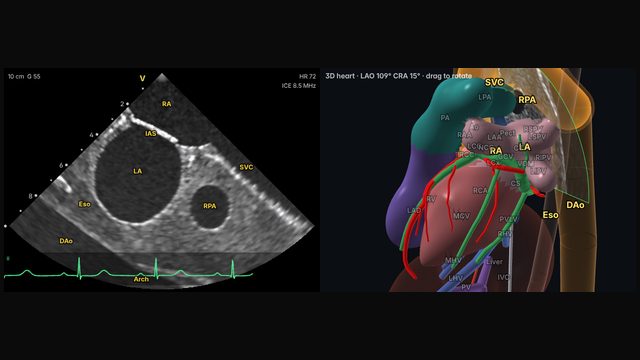
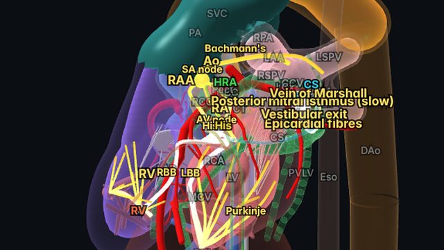
The left atrial appendage comes into view at the anterolateral edge of the left atrium, close to the mitral annulus and the left superior pulmonary vein. From the right atrium it lies in the far field, so it is seen at lower resolution than near-field structures.
Why it matters
Appendage occlusion and thrombus screening depend on seeing the appendage well. Because the right-atrial view is distant, operators often move the catheter closer for these tasks, for example into the left atrium after transseptal crossing or into the RV outflow or pulmonary artery, where the appendage sits nearer the transducer.
How to get there
About 3 o'clock (80° clockwise)
From the left atrium and mitral valve view, a little more clockwise rotation, sometimes with tilt, brings the appendage into the far field.
In the simulator:
- Start from the home view (mid right atrium, neutral).
- Rotate clockwise about 80°, to about 3 o'clock.
- Tilt left (about 20° on the R/L dial).
Clinical uses
- Guidance for left atrial appendage occlusion.
- Screening for appendage thrombus, with closer imaging positions when detail matters.
Frequently asked questions
How do you get the left atrial appendage view on ICE?
From the left atrium and mitral valve view, a little more clockwise rotation, sometimes with tilt, brings the appendage into the far field. In the Total Heart Sim model heart: Rotate clockwise about 80°, to about 3 o'clock. Tilt left (about 20° on the R/L dial). Small adjustments either way are normal: anatomy and catheters vary.
References
- Alkhouli M, Hijazi ZM, Holmes DR Jr, Rihal CS, Wiegers SE. Intracardiac echocardiography in structural heart disease interventions. JACC Cardiovasc Interv. 2018;11(21):2133–2147.
- Eleid MF, Chung CJ, Daniels MJ, et al. SCAI position statement on intracardiac echocardiography to guide structural heart disease interventions. J Soc Cardiovasc Angiogr Interv. 2025;4(12):103946.
- Enriquez A, Saenz LC, Rosso R, et al. Use of intracardiac echocardiography in interventional cardiology: working with the anatomy rather than fighting it. Circulation. 2018;137:2278–2294.
Educational content; not medical advice. Rotation and deflection values are typical teaching values in the simulator's model heart; real anatomy and catheters vary.