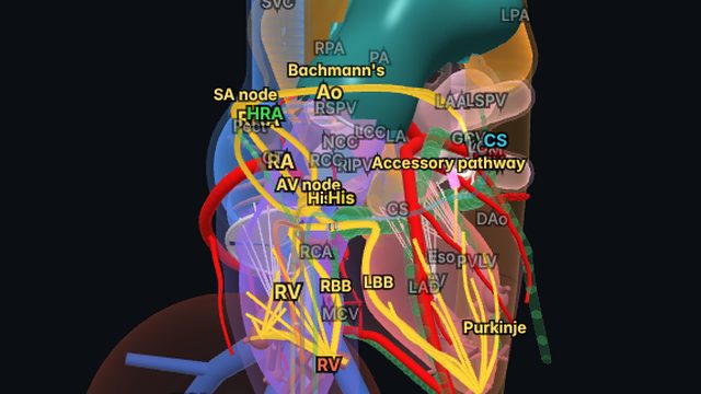
 Total Heart Sim
What you see
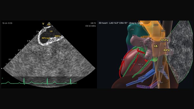
Rotating clockwise to the interatrial septum, usually with posterior deflection, lays the septum across the near field with the left atrium beyond. The fossa ovalis is the thin central part of the septum, surrounded by the thicker limbus.
Why it matters
Transseptal puncture is where ICE earns its place in many labs. The septal view shows the sheath and needle drop into the fossa, the tenting of the septum as the needle presses on it, and the distance to the posterior left atrial wall. Small rotations either side show the aortic root anteriorly and the pulmonary veins posteriorly, so the operator can choose a puncture site suited to the procedure.
How to get there
About 4 o'clock (108° clockwise)
Rotate clockwise to the interatrial septum and add posterior deflection, with a little tilt as needed, so the septum lies across the near field with the left atrium beyond.
In the simulator:
- Start from the home view (mid right atrium, neutral).
- Rotate clockwise about 108°, to about 4 o'clock.
- Deflect posteriorly (about 40° on the A/P dial).
- Tilt left (about 10° on the R/L dial).
- Set the depth to about 10 cm.
Clinical uses
- Transseptal puncture: fossa location, tenting and safe crossing.
- Choosing a puncture site for the procedure that follows.
- Assessing the septum, including patent foramen ovale and atrial septal defects.
Frequently asked questions
How do you get the septal view and fossa ovalis view on ICE?
Rotate clockwise to the interatrial septum and add posterior deflection, with a little tilt as needed, so the septum lies across the near field with the left atrium beyond. In the Total Heart Sim model heart: Rotate clockwise about 108°, to about 4 o'clock. Deflect posteriorly (about 40° on the A/P dial). Tilt left (about 10° on the R/L dial). Set the depth to about 10 cm. Small adjustments either way are normal: anatomy and catheters vary.
References
- Enriquez A, Saenz LC, Rosso R, et al. Use of intracardiac echocardiography in interventional cardiology: working with the anatomy rather than fighting it. Circulation. 2018;137:2278–2294.
- Alkhouli M, Hijazi ZM, Holmes DR Jr, Rihal CS, Wiegers SE. Intracardiac echocardiography in structural heart disease interventions. JACC Cardiovasc Interv. 2018;11(21):2133–2147.
- Eleid MF, Chung CJ, Daniels MJ, et al. SCAI position statement on intracardiac echocardiography to guide structural heart disease interventions. J Soc Cardiovasc Angiogr Interv. 2025;4(12):103946.
- Haloot J, Schurmann P. Electrophysiology laboratory survival guide part 2: introduction to intracardiac echocardiography in the electrophysiology laboratory. US Cardiology Review. 2026;20:e16.
Educational content; not medical advice. Rotation and deflection values are typical teaching values in the simulator's model heart; real anatomy and catheters vary.