 Total Heart Sim
What you see
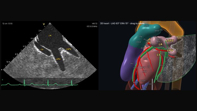
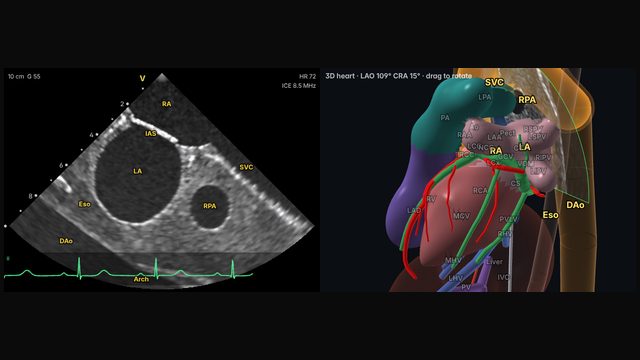
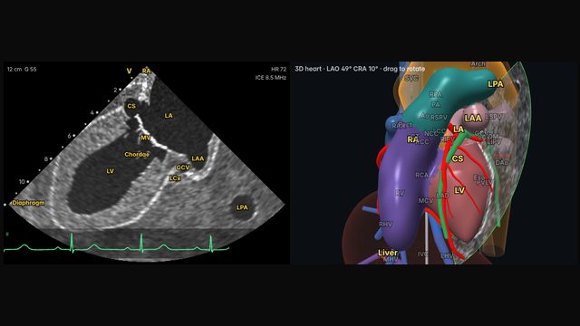
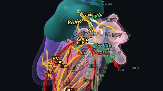
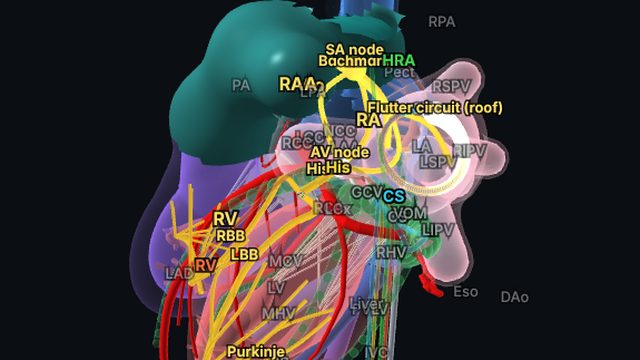
The right pulmonary veins enter the left atrium just behind the interatrial septum. Reaching them takes more rotation than the left veins, usually with posterior deflection, and the right superior and inferior veins then appear entering the posterior left atrium.
Why it matters
The right veins complete the picture for pulmonary vein isolation. Their closeness to the septum also matters for transseptal puncture: a puncture site too close to the right veins makes them awkward to reach.
How to get there
About 5 o'clock (140° clockwise)
Rotate further clockwise, often with posterior deflection, until the right superior and inferior veins appear just behind the septum.
In the simulator:
- Start from the home view (mid right atrium, neutral).
- Advance the catheter about 1.3 cm.
- Rotate clockwise about 140°, to about 5 o'clock.
- Set the depth to about 10 cm.
Clinical uses
- Pulmonary vein isolation of the right-sided veins.
- Relating the veins to the transseptal puncture site.
Frequently asked questions
How do you get the right pulmonary veins view on ICE?
Rotate further clockwise, often with posterior deflection, until the right superior and inferior veins appear just behind the septum. In the Total Heart Sim model heart: Advance the catheter about 1.3 cm. Rotate clockwise about 140°, to about 5 o'clock. Set the depth to about 10 cm. Small adjustments either way are normal: anatomy and catheters vary.
References
- Enriquez A, Saenz LC, Rosso R, et al. Use of intracardiac echocardiography in interventional cardiology: working with the anatomy rather than fighting it. Circulation. 2018;137:2278–2294.
- Haloot J, Schurmann P. Electrophysiology laboratory survival guide part 2: introduction to intracardiac echocardiography in the electrophysiology laboratory. US Cardiology Review. 2026;20:e16.
Educational content; not medical advice. Rotation and deflection values are typical teaching values in the simulator's model heart; real anatomy and catheters vary.