 Total Heart Sim
What you see
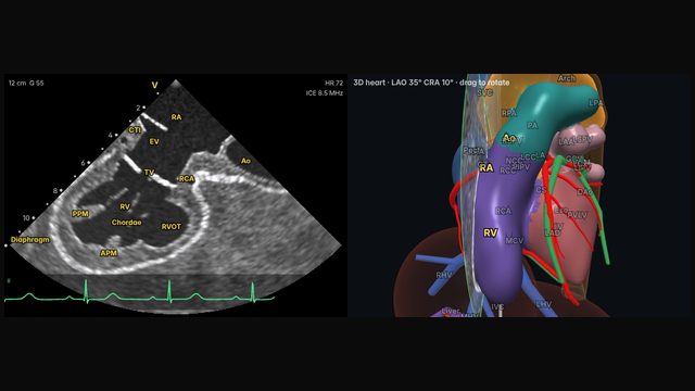
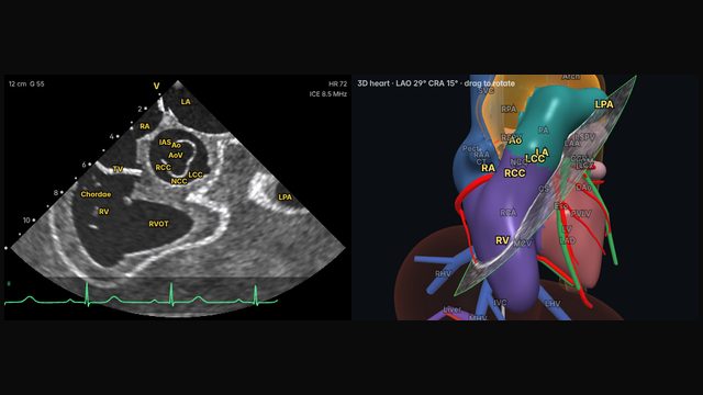
The right ventricular outflow tract runs up and to the left towards the pulmonary valve and main pulmonary artery. In this view it lies across the sector with the aortic root close behind it, which is the relationship that matters most: the RVOT wraps around the front of the aortic root and the left coronary cusp.
Why it matters
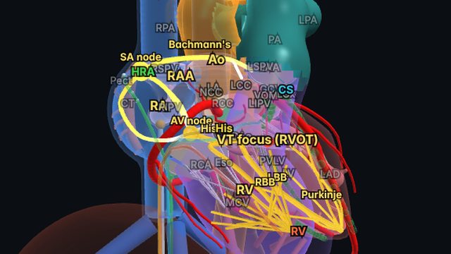
Most idiopathic ventricular arrhythmias arise in the outflow tracts. Imaging the RVOT and pulmonary valve shows where the ablation catheter sits, whether it has contact, and how close it is to the pulmonary valve and the aortic cusps behind it.
How to get there
12 o'clock, no rotation
From the home view, operators usually deflect anteriorly and advance slightly, so the plane swings up the RV outflow tract to the pulmonary valve.
In the simulator:
- Start from the home view (mid right atrium, neutral).
- Advance the catheter about 3.5 cm.
- Tilt right (about 40° on the R/L dial).
Clinical uses
- Outflow tract VT and PVC ablation: catheter position and contact.
- Pulmonary valve and main PA assessment.
Frequently asked questions
How do you get the rv outflow and pulmonary valve view on ICE?
From the home view, operators usually deflect anteriorly and advance slightly, so the plane swings up the RV outflow tract to the pulmonary valve. In the Total Heart Sim model heart: Advance the catheter about 3.5 cm. Tilt right (about 40° on the R/L dial). Small adjustments either way are normal: anatomy and catheters vary.
References
- Enriquez A, Saenz LC, Rosso R, et al. Use of intracardiac echocardiography in interventional cardiology: working with the anatomy rather than fighting it. Circulation. 2018;137:2278–2294.
- Haloot J, Schurmann P. Electrophysiology laboratory survival guide part 2: introduction to intracardiac echocardiography in the electrophysiology laboratory. US Cardiology Review. 2026;20:e16.
Educational content; not medical advice. Rotation and deflection values are typical teaching values in the simulator's model heart; real anatomy and catheters vary.