 Total Heart Sim
What it is
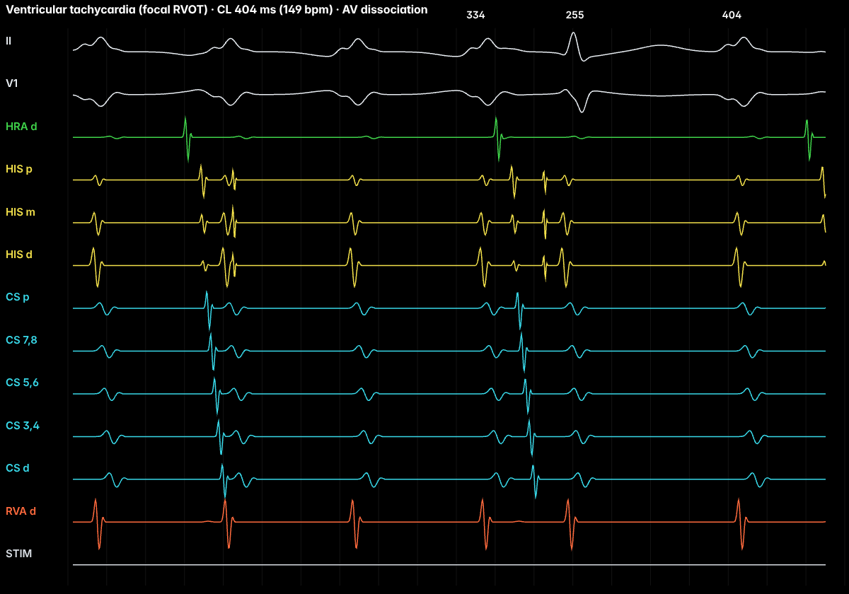
Outflow tract VT is the commonest idiopathic VT. It usually arises in the right ventricular outflow tract in a structurally normal heart and is caused by triggered activity (cAMP-mediated delayed afterdepolarizations), which is why exercise, stress and isoproterenol often provoke it and adenosine can terminate it.
On the ECG
A wide QRS with a left bundle branch block pattern and an inferior axis (tall R waves in II, III and aVF). A precordial transition at V4 or later favours the RVOT; an earlier transition raises the possibility of an LV outflow tract or aortic cusp origin. P waves may march through independently: AV dissociation, which proves VT.
In the EP lab
Burst pacing, often with isoproterenol, induces it. Activation mapping finds the earliest site, and pace mapping compares paced QRS morphology with the clinical VT or PVC.
Anatomy and ICE
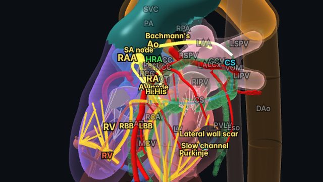
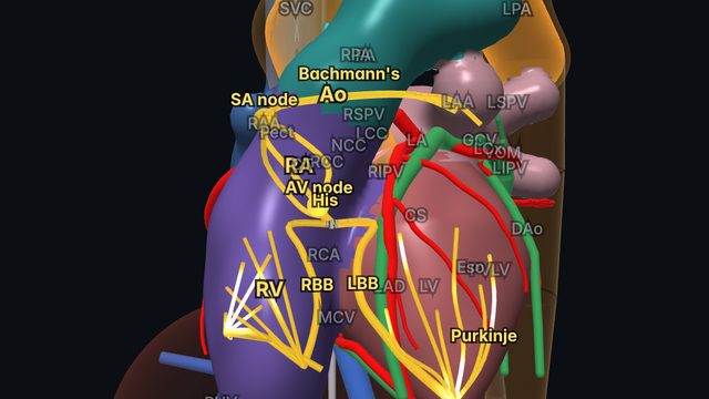
The RVOT lies just in front of the aortic root and left coronary cusp, and the pulmonary valve sits above it. ICE helps define these relationships during ablation. Arrhythmogenic right ventricular cardiomyopathy (ARVC) can produce similar morphologies and should be excluded.
Treatment and ablation
Beta-blockers or non-dihydropyridine calcium-channel blockers are options, and catheter ablation is highly effective for symptomatic outflow tract VT or frequent PVCs.
Frequently asked questions
How do you tell RVOT from LVOT origin on the ECG?
Both give an inferior axis. A precordial R/S transition at V4 or later favours the RVOT, while transition at V2 or earlier favours the LV outflow tract or aortic cusps; V3 transition is indeterminate and needs further criteria.
References
- Al-Khatib SM, et al. 2017 AHA/ACC/HRS guideline for management of patients with ventricular arrhythmias and the prevention of sudden cardiac death. Circulation. 2018;138:e272–e391.
- Zeppenfeld K, et al. 2022 ESC guidelines for the management of patients with ventricular arrhythmias and the prevention of sudden cardiac death. Eur Heart J. 2022;43:3997–4126.
- Issa ZF, Miller JM, Zipes DP. Clinical Arrhythmology and Electrophysiology: A Companion to Braunwald’s Heart Disease. Elsevier.
Educational content for clinicians and trainees; not medical advice. The simulator's rhythms are simplified models for teaching.