 Total Heart Sim
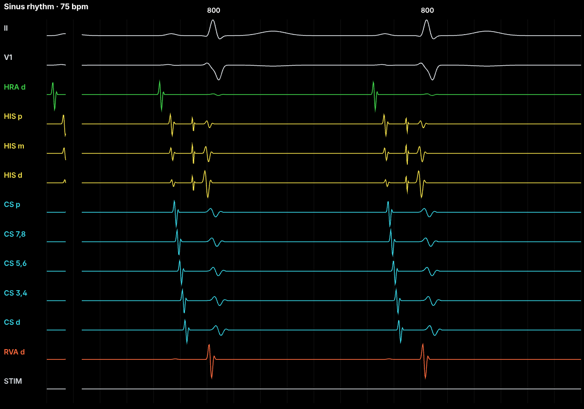
The normal sequence
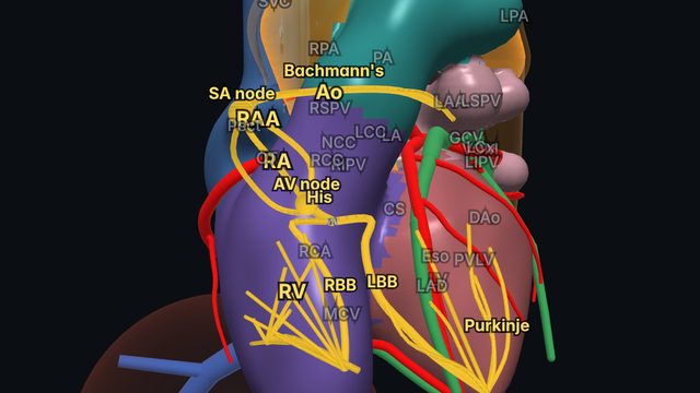
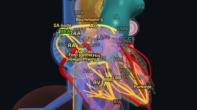
Each beat starts in the sinoatrial (SA) node near the junction of the superior vena cava and right atrium. Activation spreads across the right atrium and, through Bachmann’s bundle, to the left atrium, then slows in the AV node, which gives the ventricles time to fill. It then travels rapidly down the His bundle, the right and left bundle branches and the Purkinje network, activating the ventricles from the septum and endocardium outward.
On the ECG
Sinus rhythm has an upright P wave in leads I and II at a rate of about 60–100 bpm, a PR interval of 120–200 ms (atrial activation plus AV-nodal delay) and a narrow QRS, usually under 110–120 ms.
In the EP lab
Intracardiac recordings split the PR interval: the AH interval (AV-node conduction, roughly 50–120 ms) and the HV interval (His–Purkinje conduction, about 35–55 ms). A prolonged HV interval indicates disease below the AV node.
Anatomy and ICE
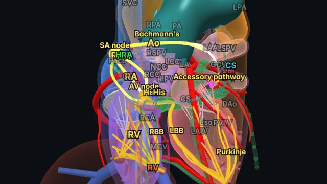
Seeing the conduction system inside the beating anatomy explains why a septal ablation risks AV block, why bundle branch block widens the QRS and how accessory pathways bypass the node. On ICE, the same beat drives the valves and walls you image.
Frequently asked questions
What are the AH and HV intervals?
Measured on the His bundle catheter, the AH interval is conduction through the AV node (about 50–120 ms) and the HV interval is conduction from the His bundle to the ventricles (about 35–55 ms). Together with atrial conduction they make up the PR interval.
References
- Josephson's Clinical Cardiac Electrophysiology: Techniques and Interpretations. Wolters Kluwer.
- Kusumoto FM, et al. 2018 ACC/AHA/HRS guideline on the evaluation and management of patients with bradycardia and cardiac conduction delay. Circulation. 2019;140:e382–e482.
Educational content for clinicians and trainees; not medical advice. The simulator's rhythms are simplified models for teaching.