 Total Heart Sim
What it is
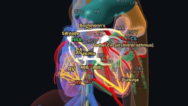
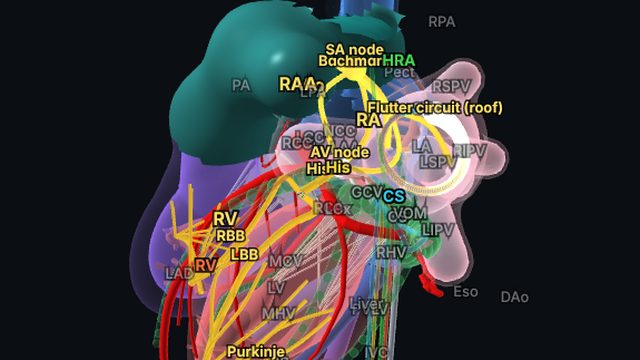
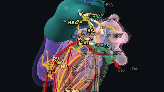
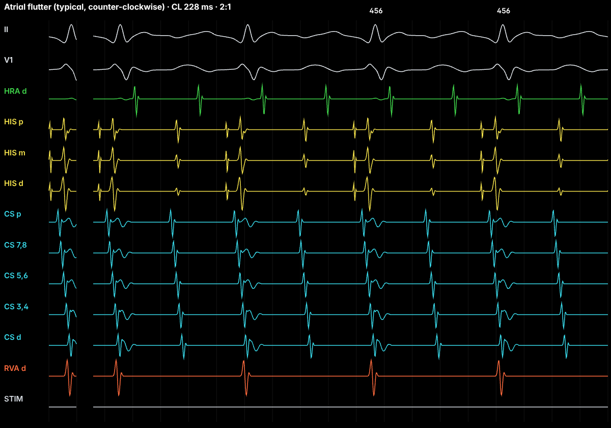
Typical atrial flutter is a macro-re-entrant circuit in the right atrium around the tricuspid annulus. Its critical, slowly conducting link is the cavotricuspid isthmus (CTI), the strip of tissue between the inferior vena cava (and Eustachian ridge) and the tricuspid valve. The usual direction is counter-clockwise viewed in LAO: up the septum, across the roof, down the lateral wall in front of the crista terminalis and through the isthmus.
On the ECG
Regular atrial activity at about 250–350 per minute with no isoelectric baseline. In counter-clockwise flutter the flutter waves are negative sawtooth waves in II, III and aVF and positive in V1; clockwise flutter shows the opposite polarity. The AV node commonly conducts 2:1, so a regular ventricular rate near 150 bpm should prompt a search for flutter waves.
In the EP lab
A 20-pole halo catheter around the tricuspid annulus shows activation marching around the ring. Entrainment from the isthmus gives concealed entrainment with a post-pacing interval close to the tachycardia cycle length (PPI − TCL within about 20–30 ms), confirming the isthmus is in the circuit. After ablation, pacing on either side of the line checks for bidirectional block.
Anatomy and ICE
The isthmus varies widely: it can be long, pouched (a sub-Eustachian recess) or crossed by a prominent Eustachian ridge, which makes lesions hard to complete. Intracardiac echo shows this anatomy and catheter contact directly, which is one reason ICE is used in flutter ablation.
Treatment and ablation
Rate control and anticoagulation follow AF principles. CTI ablation to bidirectional block is highly effective and is often first-line for recurrent typical flutter.
Frequently asked questions
Why is it called CTI-dependent?
The circuit must pass through the cavotricuspid isthmus between the IVC and the tricuspid valve. A complete ablation line across the isthmus blocks the circuit and cures the flutter.
What does bidirectional block mean?
After CTI ablation, pacing on one side of the line activates the other side only by going the long way around the annulus, in both directions. That confirms the line is complete.
References
- Page RL, et al. 2015 ACC/AHA/HRS guideline for the management of adult patients with supraventricular tachycardia. Circulation. 2016;133:e506–e574.
- Brugada J, et al. 2019 ESC guidelines for the management of patients with supraventricular tachycardia. Eur Heart J. 2020;41:655–720.
- Issa ZF, Miller JM, Zipes DP. Clinical Arrhythmology and Electrophysiology: A Companion to Braunwald’s Heart Disease. Elsevier.
Educational content for clinicians and trainees; not medical advice. The simulator's rhythms are simplified models for teaching.