 Total Heart Sim
What it is
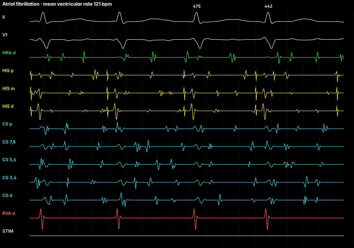
Atrial fibrillation (AF) is the commonest sustained arrhythmia. Disorganized atrial activation replaces coordinated contraction, often triggered by ectopic beats from the pulmonary veins and maintained by an atrial substrate shaped by age, hypertension, heart failure, obesity and other factors.
On the ECG
No organized P waves, a fibrillatory baseline and an irregularly irregular ventricular rhythm. The QRS is narrow unless there is aberrancy or pre-excitation.
Anatomy and ICE
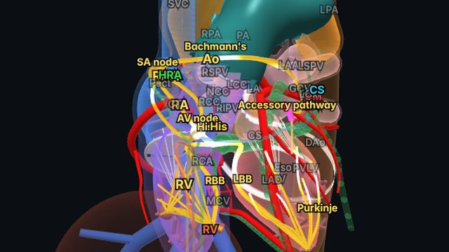
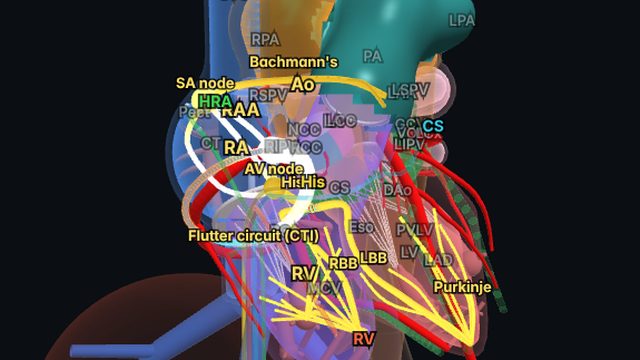
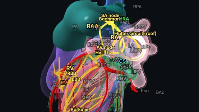
Pulmonary vein isolation is the cornerstone of AF ablation. Intracardiac echo is widely used to guide the transseptal puncture, see the pulmonary vein ostia and catheter position, monitor for pericardial effusion and locate the esophagus behind the posterior wall.
Treatment
Management combines stroke prevention (anticoagulation guided by risk scores), rate or rhythm control, risk-factor modification and, for many patients, catheter ablation, now recommended as first-line rhythm control in selected patients.
Frequently asked questions
Why is ICE used in AF ablation?
It guides the transseptal puncture, shows the pulmonary vein ostia and catheter contact, detects pericardial effusion early, and shows the esophagus relative to the posterior left atrium.
References
- Joglar JA, et al. 2023 ACC/AHA/ACCP/HRS guideline for the diagnosis and management of atrial fibrillation. Circulation. 2024;149:e1–e156.
- Issa ZF, Miller JM, Zipes DP. Clinical Arrhythmology and Electrophysiology: A Companion to Braunwald’s Heart Disease. Elsevier.
Educational content for clinicians and trainees; not medical advice. The simulator's rhythms are simplified models for teaching.