 Total Heart Sim
What it is
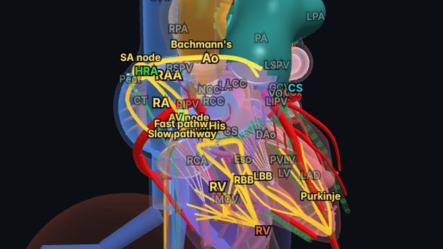
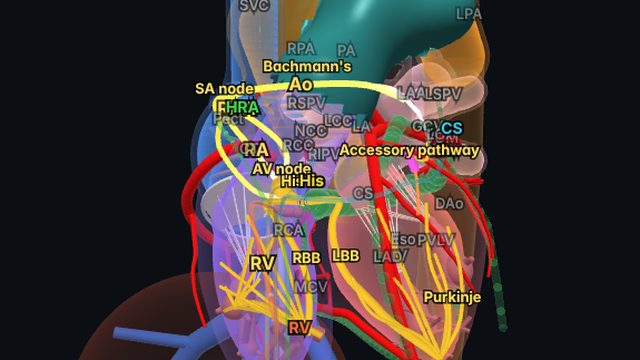
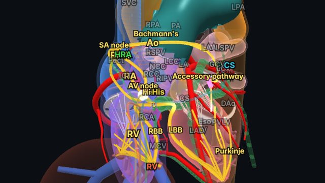
Atrioventricular re-entrant tachycardia (AVRT) uses an accessory pathway: a strand of muscle that crosses the AV groove outside the normal conduction system. In orthodromic AVRT, by far the commonest form, the impulse goes down the AV node and His–Purkinje system and back up the pathway to the atria.
A concealed pathway conducts only retrogradely (ventricle to atrium), so the resting ECG shows no pre-excitation, yet it can support orthodromic AVRT just the same.
On the ECG
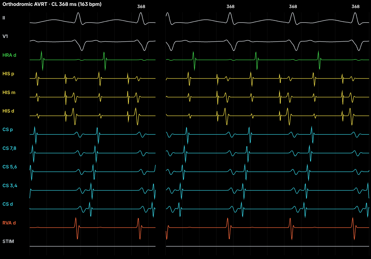
A regular narrow-QRS tachycardia (wide if there is bundle branch block). The retrograde P wave falls after the QRS, usually in the ST segment, with RP shorter than PR. Because the atria are reached through the pathway, the RP interval is longer than in typical AVNRT. Bundle branch block on the same side as the pathway lengthens the VA interval and slows the tachycardia.
In the EP lab
With a left lateral pathway, atrial activation is eccentric: earliest at the distal coronary sinus rather than the septum. Ventricular entrainment gives a V-A-V response with a short post-pacing interval (PPI − TCL usually under 115 ms), and a His-refractory PVC that advances, delays or terminates the tachycardia proves an accessory pathway participates. AV block cannot occur without terminating the tachycardia, since the ventricles are part of the circuit.
In sinus rhythm, RV pacing of a concealed left lateral pathway shows the same eccentric retrograde activation.
Anatomy and ICE
Left free-wall pathways cross the mitral annulus, which is why the coronary sinus catheter, lying along the annulus, maps them so well. Seeing the pathway, the annulus and the coronary sinus together in 3D makes the activation sequence intuitive. ICE is used to guide transseptal access to left-sided pathways and to watch catheter position on the annulus.
Treatment and ablation
Vagal manoeuvres and adenosine usually terminate it, because the AV node is in the circuit. Catheter ablation of the pathway is first-line definitive therapy for recurrent symptomatic AVRT.
Frequently asked questions
What is a concealed accessory pathway?
A pathway that conducts only from ventricle to atrium. There is no delta wave in sinus rhythm, but it can still carry orthodromic AVRT.
Why does a His-refractory PVC matter?
A PVC delivered when the His bundle is already refractory cannot reach the atria through the normal system. If it still advances or delays the next atrial activation, or terminates the tachycardia without reaching the atrium, an accessory pathway must be present.
References
- Page RL, et al. 2015 ACC/AHA/HRS guideline for the management of adult patients with supraventricular tachycardia. Circulation. 2016;133:e506–e574.
- Brugada J, et al. 2019 ESC guidelines for the management of patients with supraventricular tachycardia. Eur Heart J. 2020;41:655–720.
- Josephson's Clinical Cardiac Electrophysiology: Techniques and Interpretations. Wolters Kluwer.
Educational content for clinicians and trainees; not medical advice. The simulator's rhythms are simplified models for teaching.