 Total Heart Sim
What it is
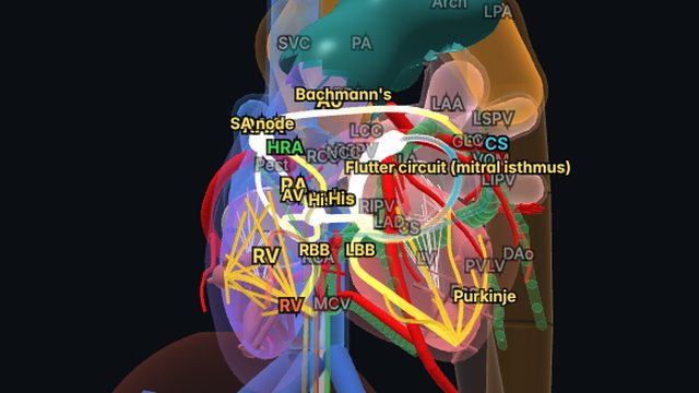
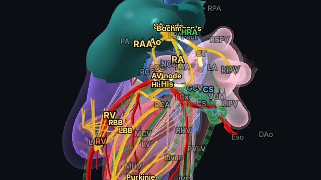
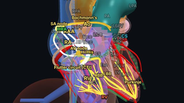
Roof-dependent flutter loops vertically around the pulmonary veins in the left atrium: up one wall, across the roof between the superior veins and down the other wall. It is common after AF ablation, where gaps in pulmonary vein isolation lines create slow conduction.
On the ECG and in the EP lab
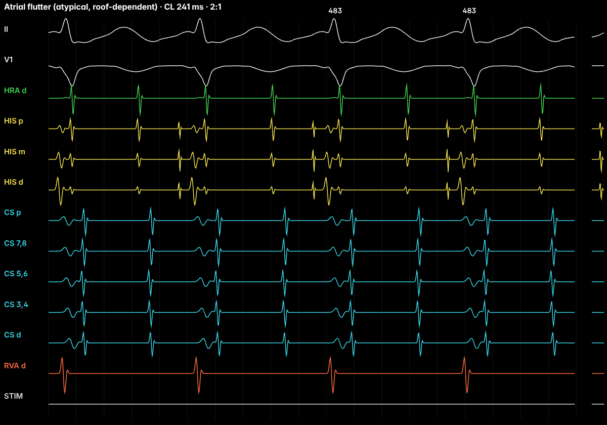
Flutter waves are low amplitude and not sawtooth. Because the wavefront travels down the anterior wall and up the posterior wall (or the reverse), the coronary sinus activates nearly simultaneously ("stacked"), and the anterior and posterior walls activate in opposite craniocaudal directions. Entrainment from the roof gives PPI − TCL near zero.
Treatment and ablation
A roof line joining the left and right superior pulmonary veins blocks the circuit. The esophagus runs behind the posterior wall, which is why the line is placed on the roof rather than low on the posterior wall.
Frequently asked questions
Why is CS activation stacked in roof flutter?
The coronary sinus runs along the inferior left atrium, perpendicular to the vertical roof circuit, so the wavefront reaches all CS electrodes at about the same time.
References
- Joglar JA, et al. 2023 ACC/AHA/ACCP/HRS guideline for the diagnosis and management of atrial fibrillation. Circulation. 2024;149:e1–e156.
- Brugada J, et al. 2019 ESC guidelines for the management of patients with supraventricular tachycardia. Eur Heart J. 2020;41:655–720.
- Issa ZF, Miller JM, Zipes DP. Clinical Arrhythmology and Electrophysiology: A Companion to Braunwald’s Heart Disease. Elsevier.
Educational content for clinicians and trainees; not medical advice. The simulator's rhythms are simplified models for teaching.