 Total Heart Sim
What it is
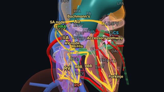
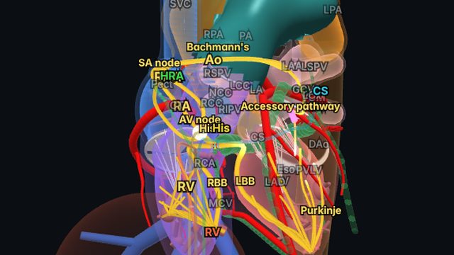
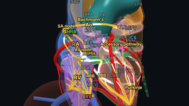
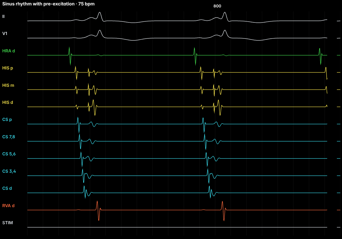
A manifest accessory pathway conducts from atrium to ventricle in sinus rhythm, bypassing the delay of the AV node. Part of the ventricle is activated early (pre-excited) through the pathway while the rest is activated normally, so each QRS is a fusion of the two. The WPW pattern describes this ECG; WPW syndrome is the pattern plus arrhythmias.
On the ECG
A short PR interval (under 120 ms), a slurred upstroke at the start of the QRS (the delta wave), a widened QRS and secondary ST–T changes. The delta-wave polarity points to the pathway: a left lateral pathway tends to give a positive delta in V1 and a negative or isoelectric delta in leads I and aVL; a posteroseptal pathway gives negative delta waves in the inferior leads that can mimic an inferior infarct.
The degree of pre-excitation varies with how quickly the AV node conducts: slowing the node (vagal tone, adenosine) increases pre-excitation, while a pathway far from the sinus node shows less.
Why it matters
Pathways can support orthodromic and antidromic AVRT, and the most dangerous situation is atrial fibrillation conducting rapidly down a pathway with a short refractory period, which can degenerate into ventricular fibrillation. Risk stratification looks at the shortest pre-excited RR interval in AF and whether pre-excitation disappears suddenly with exercise (a reassuring sign).
Anatomy and ICE
Pathways can lie anywhere around the mitral and tricuspid annuli. Septal pathways sit close to the AV node and His bundle, which raises the risk of AV block during ablation; left free-wall pathways are reached transseptally or retrogradely across the aortic valve. ICE can guide the transseptal puncture and show catheter position on the annulus.
Treatment and ablation
Catheter ablation of the pathway is recommended for symptomatic WPW syndrome and is reasonable for many people with an asymptomatic pattern after risk assessment. Success rates are high and depend on pathway location.
Frequently asked questions
What is the difference between WPW pattern and WPW syndrome?
The WPW pattern is the ECG appearance of pre-excitation (short PR, delta wave). WPW syndrome is that pattern together with symptomatic arrhythmias such as AVRT or pre-excited atrial fibrillation.
How do you localize an accessory pathway from the ECG?
From the delta-wave polarity across the 12 leads: V1 (positive suggests a left-sided pathway), leads I and aVL (negative suggests left lateral), the inferior leads (negative suggests posteroseptal or inferior) and the R/S transition. Published algorithms combine these clues.
References
- Page RL, et al. 2015 ACC/AHA/HRS guideline for the management of adult patients with supraventricular tachycardia. Circulation. 2016;133:e506–e574.
- Brugada J, et al. 2019 ESC guidelines for the management of patients with supraventricular tachycardia. Eur Heart J. 2020;41:655–720.
- Josephson's Clinical Cardiac Electrophysiology: Techniques and Interpretations. Wolters Kluwer.
Educational content for clinicians and trainees; not medical advice. The simulator's rhythms are simplified models for teaching.