 Total Heart Sim
What it is
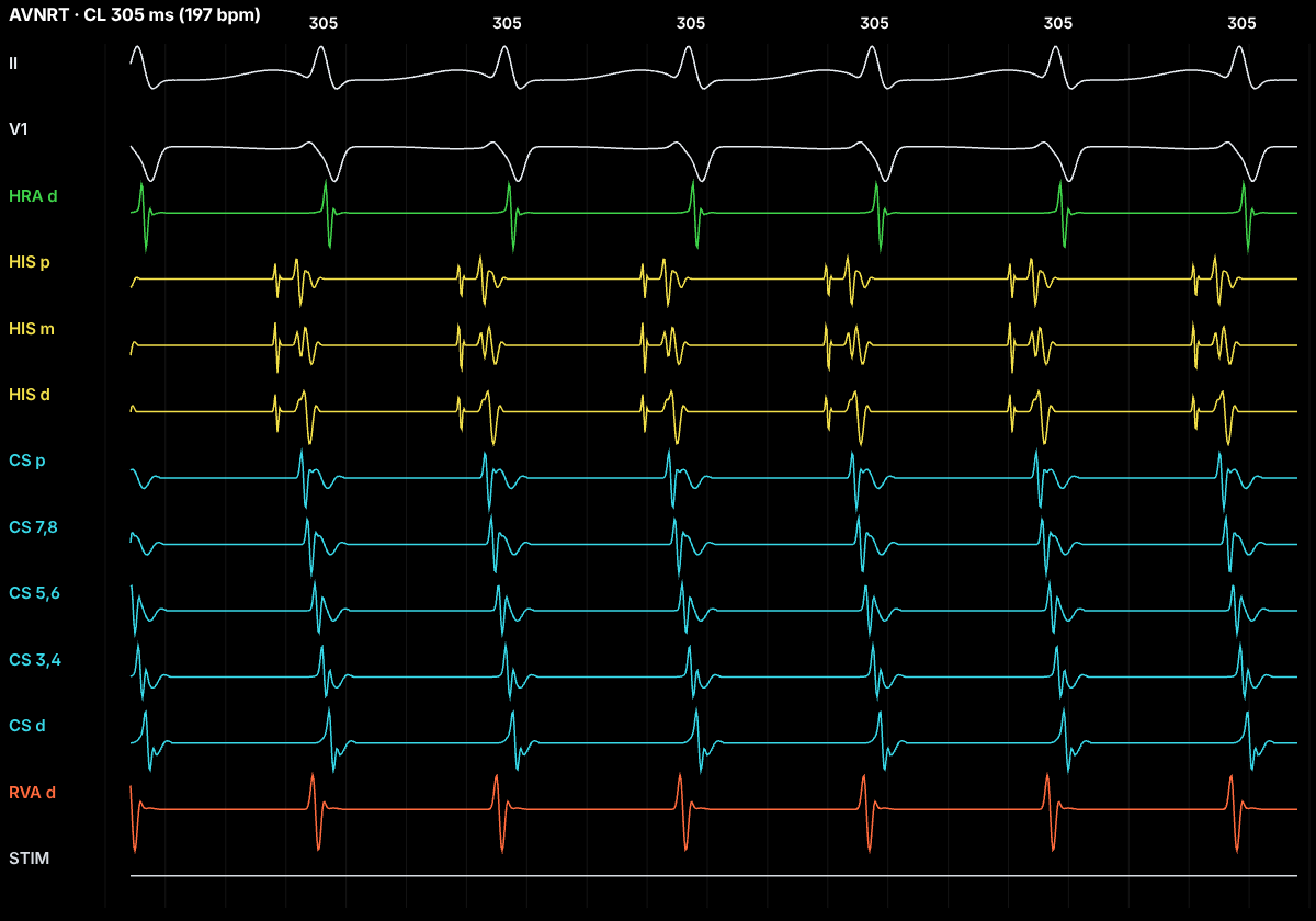
AV nodal re-entrant tachycardia (AVNRT) is the commonest regular narrow-complex tachycardia in adults after sinus tachycardia, and it is more common in women. It depends on dual AV-nodal physiology: a fast pathway with a long refractory period and a slow pathway, entering the node from near the coronary sinus ostium, with a short one.
In typical (slow–fast) AVNRT, a premature beat blocks in the fast pathway, conducts down the slow pathway, then finds the fast pathway recovered and returns up it. The impulse keeps circling, and each lap sends a wavefront down to the ventricles and up to the atria at almost the same moment.
On the ECG
A regular, narrow-QRS tachycardia, often 140–250 bpm. Because atria and ventricles activate nearly together, the retrograde P wave hides inside or just at the end of the QRS: look for a pseudo-r′ in V1 or a pseudo-S in the inferior leads that is absent in sinus rhythm.
If conduction below the circuit blocks every other beat (AVNRT with 2:1 block), the atrial rate is double the ventricular rate, with one P wave on the QRS and the next one on its own. The tachycardia continues through the block, which a circuit that needs the ventricles (AVRT) cannot do.
In the EP lab
Atrial extrastimuli reveal dual pathways as an AH jump: when the coupling interval shortens by 10 ms and the AH interval lengthens by 50 ms or more, conduction has switched from the fast to the slow pathway, and an echo beat or sustained AVNRT may follow.
During tachycardia the septal VA interval is very short (typically under 70 ms) with earliest atrial activation near the His (fast pathway). Ventricular overdrive pacing that entrains the tachycardia gives a V-A-V response with a long post-pacing interval (PPI − TCL usually over 115 ms), and a His-refractory PVC does not reset the atrium. These separate it from orthodromic AVRT and atrial tachycardia.
Anatomy and ICE
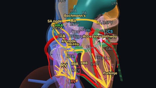
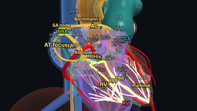
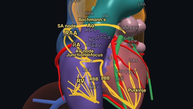
The slow pathway runs in the inferior (posterior) triangle of Koch, between the tricuspid annulus and the coronary sinus ostium; the compact AV node and His bundle sit at the apex of the triangle. Seeing these structures in 3D explains why slow-pathway ablation is done low and posterior, away from the His.
On intracardiac echo the coronary sinus ostium, the tricuspid annulus and the septal region of the triangle of Koch can be imaged, which helps place and stabilise the ablation catheter.
Treatment and ablation
Vagal manoeuvres or adenosine usually terminate it acutely, because the AV node is part of the circuit. Catheter ablation (slow-pathway modification) is first-line definitive therapy for recurrent symptomatic AVNRT, with high success and a small risk of AV block.
Frequently asked questions
How do you tell AVNRT from orthodromic AVRT?
In typical AVNRT the VA interval is very short (septal VA under about 70 ms, P wave hidden in the QRS), atrial activation is concentric, ventricular entrainment gives PPI − TCL over about 115 ms, and a His-refractory PVC does not reset the atrium. In orthodromic AVRT the VA interval is longer, activation is often eccentric, PPI − TCL is shorter and a His-refractory PVC that resets the atrium proves an accessory pathway.
What is an AH jump?
An increase in the AH interval of 50 ms or more when the atrial extrastimulus coupling interval shortens by 10 ms. It marks block in the fast AV-nodal pathway and conduction down the slow pathway: the electrophysiological signature of dual AV-nodal physiology.
Can AVNRT continue with AV block?
Yes. The circuit lies in the AV node and the atrial tissue around it, so block below the circuit (2:1 AVNRT) does not stop it. AV block during tachycardia excludes orthodromic AVRT, which needs the ventricles.
References
- Page RL, et al. 2015 ACC/AHA/HRS guideline for the management of adult patients with supraventricular tachycardia. Circulation. 2016;133:e506–e574.
- Brugada J, et al. 2019 ESC guidelines for the management of patients with supraventricular tachycardia. Eur Heart J. 2020;41:655–720.
- Josephson's Clinical Cardiac Electrophysiology: Techniques and Interpretations. Wolters Kluwer.
Educational content for clinicians and trainees; not medical advice. The simulator's rhythms are simplified models for teaching.