 Total Heart Sim

What it is
Junctional ectopic tachycardia comes from abnormal automaticity in the AV junction (the AV node and His bundle). The ventricles are activated through the normal His–Purkinje system, while the atria are activated retrogradely, or not at all if there is retrograde block.
It is uncommon in adults, where it may follow cardiac surgery or occur with digoxin toxicity, myocarditis or high catecholamine states. It is best known in children after congenital heart surgery and as a rare congenital form. An accelerated junctional rhythm is also commonly seen during slow-pathway ablation for AVNRT, where it marks heating near the slow pathway.
On the ECG
A narrow QRS identical to the sinus QRS, at a rate faster than the sinus rate. Retrograde P waves, inverted in the inferior leads, appear just after the QRS, or the atria and ventricles dissociate.
Anatomy and ICE
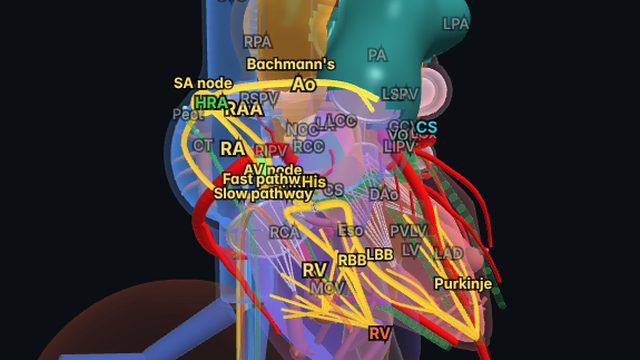
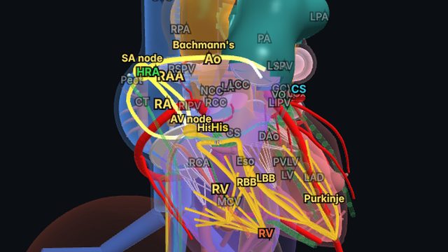
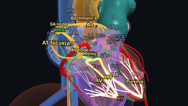
The focus sits at the apex of the triangle of Koch, where the compact AV node continues into the His bundle at the membranous septum. In 3D, the impulse can be followed down the bundle branches and back up into the atria.
Treatment
Treatment targets the cause: stopping digoxin, correcting electrolytes, reducing catecholamines. Beta-blockers and amiodarone are used, and children after surgery may also be cooled and paced. Ablation near the AV node carries a risk of AV block and is reserved for refractory cases.
Frequently asked questions
How is junctional tachycardia different from AVNRT?
Junctional tachycardia is automatic: it is not started or stopped by pacing and can continue with VA block or AV dissociation. AVNRT is re-entrant: extrastimuli start it, pacing can entrain and terminate it, and it usually has 1:1 VA conduction.
References
- Page RL, et al. 2015 ACC/AHA/HRS guideline for the management of adult patients with supraventricular tachycardia. Circulation. 2016;133:e506–e574.
- Brugada J, et al. 2019 ESC guidelines for the management of patients with supraventricular tachycardia. Eur Heart J. 2020;41:655–720.
- Issa ZF, Miller JM, Zipes DP. Clinical Arrhythmology and Electrophysiology: A Companion to Braunwald’s Heart Disease. Elsevier.
Educational content for clinicians and trainees; not medical advice. The simulator's rhythms are simplified models for teaching.